Why Should You Choose a 3D Live Cell Imaging Microscope for Advanced Research?
2025-09-09
Modern biological and medical research relies heavily on accurate visualization of living cells. Traditional two-dimensional imaging often fails to capture the complexity of dynamic cellular processes. This is where the 3D Live Cell Imaging Microscope comes in—an advanced scientific instrument designed to provide precise, real-time imaging of living cells in three dimensions.
What Is a 3D Live Cell Imaging Microscope?
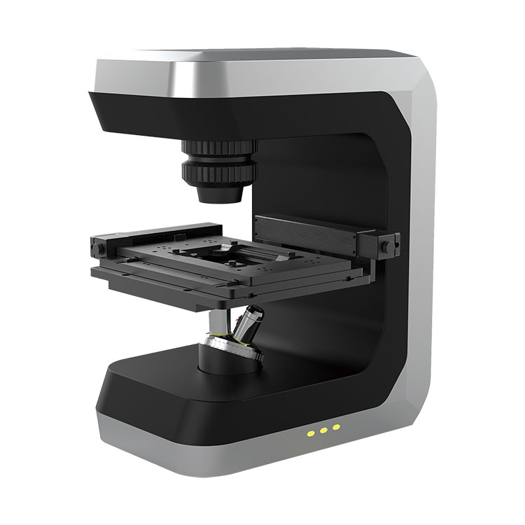
A 3D Live Cell Imaging Microscope is a cutting-edge optical system engineered to monitor cellular structures and processes in their natural environment. Unlike conventional microscopes, it allows researchers to observe cells in real time without causing damage, ensuring that the biological activity remains intact. This makes it an indispensable tool for life science laboratories, pharmaceutical development, and clinical research institutions.
Key Features and Parameters
Below are the main parameters of our 3D Live Cell Imaging Microscope, developed and supplied by Bojiong (Shanghai) Precision Machinery Technology Co., Ltd.:
-
Imaging Dimension: 2D and 3D live imaging
-
Resolution: Up to 120 nm lateral and 350 nm axial
-
Objective Lenses: 20x, 40x, 60x oil/water immersion options
-
Fluorescence Channels: Up to 6 channels (UV, Blue, Green, Red, Far Red, Infrared)
-
Live Cell Chamber: Controlled environment with temperature (25–40°C), humidity, and CO₂ regulation
-
Imaging Speed: 30 frames per second for real-time observation
-
Data Output: High-resolution TIFF, RAW, and 3D reconstruction formats
-
Software: Intuitive image analysis with automated quantification
Product Parameters in Table Format
| Parameter | Specification |
|---|---|
| Imaging Modes | 2D & 3D live cell imaging |
| Resolution | 120 nm (lateral), 350 nm (axial) |
| Objective Lenses | 20x, 40x, 60x (oil/water immersion) |
| Fluorescence Channels | 6 (UV, Blue, Green, Red, Far Red, Infrared) |
| Environmental Control | Temperature: 25–40°C, Humidity: 90%, CO₂: adjustable |
| Frame Rate | Up to 30 fps |
| Data Export | TIFF, RAW, 3D reconstruction formats |
| Software | Automated analysis, cell tracking, quantitative data reports |
Benefits of Using a 3D Live Cell Imaging Microscope
-
Real-Time Observation – Capture living cells as they move, divide, and interact.
-
High Resolution – Gain detailed visualization of intracellular structures.
-
Non-Invasive Imaging – Cells remain alive and unaffected during observation.
-
Comprehensive Data – Obtain quantitative results for in-depth analysis.
-
Versatile Applications – Suitable for drug discovery, cancer research, immunology, and neuroscience.
Applications in Research and Industry
-
Drug Discovery: Screen compounds and monitor cellular responses in real time.
-
Cancer Research: Track tumor cell migration and drug sensitivity.
-
Stem Cell Biology: Observe stem cell differentiation and regeneration processes.
-
Immunology: Visualize immune cell activity and interactions.
-
Neuroscience: Explore neuronal networks and synaptic activity.
Frequently Asked Questions (FAQ)
Q1: What makes a 3D Live Cell Imaging Microscope different from a conventional microscope?
A1: Unlike traditional microscopes, a 3D Live Cell Imaging Microscope allows real-time three-dimensional visualization of living cells. It integrates advanced fluorescence imaging and environmental control, ensuring cells remain alive during extended observation.
Q2: Can the system handle multiple fluorescence channels at once?
A2: Yes, the microscope supports up to six fluorescence channels, enabling simultaneous multi-color imaging. This allows researchers to label and track different cellular components in one experiment.
Q3: Is the imaging process harmful to living cells?
A3: No, the system uses optimized illumination and environmental control to minimize phototoxicity. Cells remain viable, allowing researchers to study natural biological processes without interference.
Q4: What industries or fields benefit most from using this technology?
A4: Life science laboratories, pharmaceutical companies, hospitals, and universities benefit significantly. Applications include cancer biology, regenerative medicine, neuroscience, immunology, and drug development.
Why Choose Bojiong (Shanghai) Precision Machinery Technology Co., Ltd.?
With years of expertise in precision optical engineering, Bojiong (Shanghai) Precision Machinery Technology Co., Ltd. delivers reliable, high-performance imaging systems tailored to advanced research needs. Our 3D Live Cell Imaging Microscope combines innovation, durability, and intuitive usability, making it the perfect choice for professionals seeking accurate and long-lasting results.
Conclusion
The 3D Live Cell Imaging Microscope is revolutionizing the way scientists study living cells. With high-resolution imaging, advanced environmental control, and powerful analytical software, it has become an essential tool for modern research. Whether you are advancing drug discovery, studying complex diseases, or exploring cellular biology, this microscope ensures precision and reliability.
For more information or to request a detailed quotation, please contact Bojiong (Shanghai) Precision Machinery Technology Co., Ltd. and discover how our advanced imaging solutions can support your research goals.


